
During the very first weeks of pregnancy, the embryo does not yet receive maternal blood. It draws its resources from a small structure called the yolk sac, somewhat like an onboard reserve. Establishing a true exchange circuit between the mother and the future baby takes time, and each step is important.
The yolk sac: the first source of nutrients before the placenta
Before the placenta takes over, the embryo relies on a temporary structure. The yolk sac provides the first nutrients and participates in the formation of the first blood cells. This system remains active for several weeks.
Wondering why the placenta doesn’t intervene right away? Because it doesn’t exist yet. Its construction begins as soon as implantation occurs, but it takes time for it to become functional. Understanding when maternal-fetal blood exchange begins requires distinguishing between these two phases: the autonomous phase (yolk sac) and the connected phase (placenta).
During this period, the embryo grows without directly receiving maternal blood. The yolk sac ensures the transition until the placental vascular network is ready to function.
Implantation and lacunar stage: the very first blood contacts
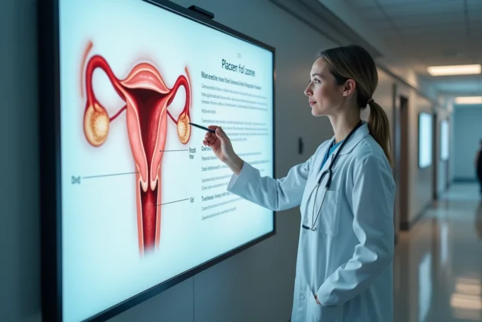
Implantation occurs about six to ten days after fertilization. The embryo, now a blastocyst, implants into the wall of the uterus. At this stage, a layer of cells called the trophoblast begins to invade the endometrium (the uterine lining).
This is where things become concrete. The trophoblast divides into two parts. One of them, the syncytiotrophoblast, digs small cavities in the maternal tissue. These cavities have a specific name: lacunae.
Lacunae and maternal blood: contact without mixing
Between the 9th and 12th day after fertilization, these lacunae gradually fill with maternal blood. They communicate with the vessels of the endometrium. This is the very first contact between the mother’s blood and the embryonic tissues.
A key point to remember: there is never a direct mixing of the two bloods. The maternal blood and fetal blood do not touch. Exchanges occur through a thin membrane, the placental barrier, which filters substances. Oxygen and nutrients pass one way, while carbon dioxide and waste products go the other way.
Imagine a very thin partition between two rooms. Air circulates through, but the occupants of each room never cross paths. This is exactly how the placental barrier works.
Functional uteroplacental circulation: the real turning point of the first trimester
The lacunar stage initiates minimal exchange. The fully operational placental circulation sets in later. Embryology data places the onset of uteroplacental flow around the 7th week of pregnancy.
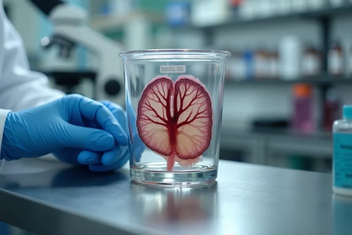
From that moment on, the fetal tissue receives oxygen and nutrients via the vessels of the umbilical cord. The placenta then becomes the central organ of pregnancy. It performs several simultaneous functions:
- The transfer of oxygen from maternal blood to fetal blood, and the removal of carbon dioxide in the opposite direction
- The supply of nutrients (glucose, amino acids, vitamins) necessary for the development of the fetus’s organs
- The production of hormones that maintain the pregnancy and prepare the mother’s body for childbirth
- The role of a protective filter, blocking some infectious agents and toxic substances
The umbilical cord connects the fetus to the placenta. It contains two arteries and one vein. The arteries carry deoxygenated blood from the fetus to the placenta, where it is recharged with oxygen through (indirect) contact with maternal blood. The vein then returns the oxygenated blood to the fetus.
Placental barrier: why the mother and fetus do not share their blood
This point deserves attention, as it is often misunderstood. Many expectant parents imagine that the mother’s blood flows directly into the baby’s body. This is not the case.
The placental barrier physically separates the two blood circulations. It consists of several layers of cells that allow certain molecules to pass through by diffusion or active transport, while blocking others.
This barrier is not perfect. Some substances still cross it: alcohol, nicotine, certain medications, and some infectious agents. This is why health recommendations during pregnancy emphasize dietary and medication precautions so strongly.
Fetal microchimerism: cells that cross the barrier
A lesser-known phenomenon deserves mention. During pregnancy, small amounts of fetal cells cross the placental barrier and enter the maternal circulation. These cells can persist in the mother’s body for years after birth. This phenomenon is called fetal microchimerism.
This is not a blood exchange per se, but a cellular transfer. Research on this topic continues to explore its implications for long-term maternal health.
Summary of key stages of maternal-fetal exchange
| Period | What happens |
|---|---|
| Day 6 to 10 after fertilization | Implantation of the blastocyst in the uterine wall |
| Day 9 to 12 | Lacunar stage: first contact between lacunae and maternal blood |
| Weeks 3 to 6 | Progressive construction of the placenta, yolk sac still active |
| Around the 7th week | Functional uteroplacental flow, the placenta takes over |
| End of the 1st trimester | Fully effective placental circulation, exchanges at full speed |
The blood exchange between the mother and the fetus does not activate in an instant. It builds up in stages, from the first lacunae filled with maternal blood to the complete placental circulation by the end of the first trimester. Maternal blood and fetal blood remain separate throughout the pregnancy, connected only by this filtering membrane that nourishes, protects, and regulates.